HOW CAN WE HELP YOU? Call 1-800-TRY-CHOP

In This Section
LiBI Gathers Experts to Explore How Early Life Influences Neurodevelopment
Published on April 12, 2019 in Cornerstone Blog · Last updated 1 month 1 week ago
 By Jillian Rose Lim, Barbara Drosey, and Nancy McCann
From discussions on early-life adversity to autism spectrum disorder (ASD), the Lifespan Brain Institute (LiBI) delivered a stimulating second symposium April 8, gathering experts across diverse disciplines to share novel research into how brain and behavior develop over a lifetime. Two hundred attendees learned about cutting-edge basic and translational research projects that address a complex and critical question: What are the factors in early life that place some individuals at risk for neuropsychiatric disorders, while others are resilient?
LiBI is a unique collaboration between Children’s Hospital of Philadelphia and the University of Pennsylvania that supports research across the fetal-adult continuum, a pillar of CHOP Research Institute’s strategic plan. By bridging the gap between pediatric and adult research, LiBI’s “lifespan approach” enables researchers to begin studying participants at a young age and to continue to follow them into adulthood.
“This idea that we would have the opportunity to understand mental health conditions starting at birth was really novel,” said Tami Benton, MD, psychiatrist-in-chief at CHOP in her opening remarks about LiBI. “It’s been an amazing journey to see the birth of this idea translate into the recruitment of new scientists, new research publications, new findings, and truly energize the research enterprise. This has become an incredible vision.”
LiBI’s Director, Raquel Gur, MD, PhD, professor of Psychiatry, Neurology, and Radiology at Penn, described LiBI’s ongoing mission in her introductory remarks: “The dream is that in the future, we will be able to reliably identify in young people — as young as we can go — those who are at increased risk for developing significant brain abnormalities, and we will be able to tweak it so that we can maximize functioning as young as children and young adults.”
Adversity and Anhedonia in Early Life: Opening Keynote
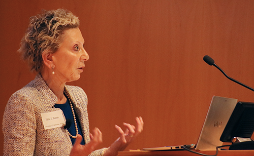 Tallie Z. Baram, MD, PhD, director of the Conte Center at the University of California Irvine, kick-started the day with a keynote that sparked excitement and discussion in the audience. The presentation focused on research from Dr. Baram’s lab that investigates the ways in which early-life experiences can disrupt brain circuit maturation and promote vulnerability to neuropsychiatric disorders.
Throughout prenatal life, the number of neurons that grow, divide, and organize is immense. During this time of significant vulnerability, an individual’s brain circuits are immature, and synaptic connections are malleable. To show how critical this window of time can be, Dr. Baram highlighted findings from an animal study in which exposure to just one week of simulated poverty (reduced bedding and nesting in a rat family’s cage) was associated with cognitive problems and progressive deficits in memory in the rat pups as adolescents. The rats exposed to early-life adversity also showed a reduced ability to experience pleasure, a concept known as anhedonia. Dr. Baram explained that anhedonia is an important focus because it indicates dysfunction in the brain’s reward circuitry.
“The question remains, what is the connection between adverse early experiences and changes in the structure of the brain?” Dr. Baram said. “What are the signals that adversity might emit to disrupt the normal maturation of brain cells?”
The answer may lie in the sensory signals that a mother imparts to her infants in early life, she continued, as her findings suggest that unpredictable and fragmented sensory signals from a rodent mother influenced pups’ emotional disorders later in life.
Novel Genes and Neurological Differences in Children
Elizabeth Bhoj, MD, PhD, clinical geneticist at CHOP, followed the keynote with a fascinating glimpse into how researchers are working toward identifying novel genes and pathways that cause neurological differences in children with craniofacial and neurodevelopmental abnormalities.
“Craniofacial anomalies can be used as a marker that [children] are more likely to have a single-gene cause,” said Dr. Bhoj, who presented several research projects relevant to LiBI.
One such breakthrough was her identification of two sisters with macrocephaly who also shared mutations in the TBC1 domain-containing kinase (TBCK) gene. Dr. Bhoj showed that the sisters had a novel pediatric neurodevelopmental syndrome, TBCK-related encephalopathy, that was co-discovered by Dr. Bhoj and colleagues in the Center for Applied Genomics at CHOP.
“Because it’s clearly degenerative, we have a window to intervene,” Dr. Bhoj said.
Her team is studying the effectiveness of leucine supplements in children identified with TBCK-related ID syndrome as a potential treatment.
 Encouraging New Neuron Development
Amelia Eisch, PhD, scientist in the Department of Anesthesiology and Critical Care Medicine at CHOP and professor of Anesthesiology and Critical Care at Perelman School of Medicine at the University of Pennsylvania, presented a whirlwind of information in “New neurons in the postnatal brain: What are they good for? And what is good for them?”
“The translational relevance opportunities here [at CHOP and Penn] are great,” Dr. Eisch said, as she summarized current collaborative lab projects involving the inducible activation and silencing of new neurons for effects in neonatal abstinence syndrome, traumatic brain injury, and perinatal brain injury.
Providing a “30,000 foot view” of her and colleagues’ volume of work, Dr. Eisch focused on her NIH-funded project investigating the dentate gyrus (DG) and its effect on memory, mood regulation, pattern separation, and reward.
So, what are new neurons in the postnatal brain good for? Quite a bit, it seems. When neurogenesis is reduced in an animal model, diminished hippocampal function results in less proficiency in spatial learning; more signs of depression in times of stress; less responsiveness to antidepressants; and a buffered sensitivity of reward circuits.
As for what’s good for neurogenesis? “Learning, exercise, and an enriched environment,” Dr. Eisch said.
DG function is disrupted in many psychiatric disorders, so part of Dr. Eisch’s work involves how to normalize or improve DG function by stimulating the entorhinal cortex-DG pathway, which has been shown to aid memory task performance, normalize mood, and improve pattern separation skills.
“While we try to understand the abnormal brain, we’re also learning about the baseline brain,” said Dr. Eisch, whose takeaway for attendees was, “Neurogenesis is a process, not a time point.”
Maternal-Fetal Health
Tamar Gur, MD, PhD, assistant professor, Department of Psychiatry, Ohio State University, Institute for Behavioral Medicine Research and Perelman School of Medicine alumna, presented “The Effects of Prenatal Stress of Fetal Brain.” In her work, Dr. Gur found that stress and mental illness are an overlapping phenomenon, with inflammation being a common point between the two.
Dr. Gur’s team investigates cytokines capable of entering fetal circulation; the hypothalamic-pituitary-adrenal (HPA) axis and the corticotropin-releasing hormone that influences the developing fetus; epigenetics to examine stress-modified gene expression; and microbiota, which she described a new frontier. One aspect of her research is HPA activation and its effect on microbiota and the intestinal epithelium.
“The uterus is close to the gut and highly vascularized,” Dr. Gur said. “It’s not a far stretch to think how these things can affect one another.”
Working with a mouse model, Dr. Gur and her colleagues demonstrated that stress during gestation leads to a shift in microbes in the dam that trigger an immune response via increased CCL2, IL-β, IL-6, and toll-like receptor 4 (TLR4); increased inflammation; and social behavior effects. Mice birthed from the stressed dam had increased IL-1β and IL-6 in adulthood, demonstrating that what affected the fetus was carried through to the adult brain.
Next, Dr. Gur and her team wondered if the increase in cytokines was microbe dependent, so they administered antibiotics to dams exposed to stress. In brief, mice that received antibiotics showed no increase in CCL2, IL-6, or TLR4, with behavioral effects to be determined. This finding suggests that influencing the microbiome during pregnancy may benefit both mother and offspring.
3D Organization of Genomes
Jennifer Phillips-Cremins, PhD, assistant professor of bioengineering and New York Stem Cell Foundation-Robertson Investigator, Perelman School of Medicine at the University of Pennsylvania, shared her work in “3D Epigenome Reconfiguration in Brain Development and Disease.” She asked attendees to think of the genome, folded into a hierarchy of topologically associating domains (TADs) and subTADs, as a series of Russian nesting dolls.
“Our genome that encodes all the functions of all our cells, and could be stretched past 6 feet, folds into complicated and intricate patterns that fit on the head of a pin,” Dr. Phillips-Cremins said.
Her lab team at the Penn Epigenetics Institute is exploring the role of the boundaries between the “dolls” and genome misfolding in trinucleotide repeat expansion disorders such as Fragile X syndrome. While there are approximately 30 diverse unstable repeat expansion disorders, the repeated sequence and mutation length vary widely among them.
Could her team find a unifying principle to find why certain genes become unstable and others do not? They hypothesized that unique 3D genome folding patterns might occur at unstable short tandem repeats (STRs). They devised seven statistical tests, with all results demonstrating disease-associated STRs were found at the boundary of TADs more than their unexpanded counterparts. Additionally, the team found a two-part predictive model for unstable expansion: boundaries enriched with the architectural protein CTCF that also had a high density of CpG islands (regions of the genome that contain a large number of CpG dinucleotide repeats).
In her self-described “second most exciting day in the lab,” Dr. Phillips-Cremins and her team discovered that the boundary co-localized with FMR1 disease-associated STR is structurally disrupted in Fragile X syndrome, silencing FMR1. Next steps include follow-up studies to assess cause and effect between FMR1 silencing and boundary integrity.
“We hope to improve and control cellular function with technologies we’re developing to manipulate genome structure,” Dr. Phillips-Cremins said.
Biomarkers for Autism Spectrum Disorder
During “Multimodal Imaging/Electrophysiologic Studies of Autism Spectrum Disorder (ASD): From Children to Adults,” Timothy Roberts, PhD, vice-chair of research for the Department of Radiology and the Oberkircher Family Endowed Chair in Pediatric Radiology at CHOP, payed homage to the “big machines”— imaging techniques such as MRI, magnetic resonance spectroscopy, and magnetoencephalography (MEG), diffuser tensor imaging, and high-angular resolution diffusion imaging — that are helping provide neurochemical insights and enhanced understanding of the autistic brain across the lifespan.
For example, a one-of-a-kind MEG machine at CHOP, designed for infants to children 3 years of age, enables effective evaluation of the smallest patients for evoked responses, which could be an early indicator of an ASD diagnosis with ramifications for early intervention.
Dr. Roberts, also a professor of radiology at Perelman School of Medicine, highlighted his research in pursuit of biomarkers for diagnosis and prognosis, as well as stratification, of ASD. More than a decade ago, researchers observed a delay in evoked response to auditory stimulation in children with ASD. Dr. Roberts’ work with latency of auditory-evoked M100 and delayed change detection of vowel categorization contributed to the correlation of M100 latency with ASD.
When evoking a response to vowel sounds (e.g., /a/,/a/,/a/,/u/), latency of the vowel-change detection was most prevalent in children with a vocabulary comprised of fewer than 30 words. In follow-up with the evoked-response subjects, researchers were able to predict the child’s behavioral and clinical state from the original data, demonstrating that M100 has prognostic value. Additionally, his team has identified gamma band activity as another component of the evoked response.
“By studying gamma and GABA (gamma-aminobutyric acid) in the same individuals through development, we found persistent gamma deficits in adults with ASD,” Dr. Roberts said, noting a coupling of gamma and GABA in children appears to be necessary for optimal formation of brain circuitry.
Measuring Brain Activity in Neurodevelopmental Disorders
J. Christopher Edgar, PhD, director of the Brains Change Pediatric Brain Imaging Program at CHOP and associate professor of Radiology in the Perelman School of Medicine at the University of Penn, enlightened the symposium attendees with his talk, “Identifying Electrophysiology Biomarkers Against a Backdrop of Normal Brain Development: Autism Spectrum Disorder and Schizophrenia.” He reviewed studies demonstrating that brain measures showing abnormal brain activity in individuals with neurodevelopmental disorders, such as ASD or schizophrenia, are of clinical use only for specific age ranges.
“It is hypothesized that children with ASD will show a different maturation rate of brain rhythms and patterns than controls, with rate of change rather than singe time-point measures better separating ASD and controls,” Dr. Edgar said, and that, “rate of change rather than single time-point measures will also better predict symptom severity.”
In a more recent Brains Change study, Dr. Edgar’s team obtained measures of brain function and structure from infancy to toddlerhood to understand normal brain development. Read about Dr. Edgar’s new federally funded infant study “Brain Structure and Function in Infants,” in his recent Q&A on Cornerstone.
Cognitive Control, Autism, and Attention-deficit/Hyperactivity Disorder
“Autism is like a vise on your brain. And each unexpected event is like another turn on the vise … it just keeps building until you feel like you’re going to explode. Sometimes when you explode, it comes out the wrong way.”
Using this quote from a young student who has autism as the opening for his talk “Cognitive Control in Autism: From Brain to Outcomes,” Benjamin Yerys, PhD, a psychologist in the Department of Child and Adolescent Psychiatry and Behavioral Sciences at CHOP, wowed his audience right from the start.
Highlighting recent and past research, Dr. Yerys demonstrated why cognitive control matters for autism and how it relates to outcomes. Past research took a simpler view — looking at single brain areas. But, according to Dr. Yerys, it’s so much more than that.
“It’s about dynamic activity between multiple brain areas within a network and between multiple networks that allow us to meet the demands of a cognitive control task,” he said
The overlay of autism spectrum disorder and attention-deficit/hyperactivity disorder is another area of research Dr. Yerys covered. He noted it’s now becoming more accepted that some cognitive control impairments may be related to a second diagnosis of ADHD, and not to the core of autism. Cognitive control affects both daily functioning and also response to treatment. When doctors try to treat ADHD with medications, they tend to be far less successful in children with autism.
Dr. Yerys sees promise in a novel digital treatment called Project: Evo, of which he is a consultant to the manufacturer. The premise behind the treatment is that cognitive control can be improved by enhancing the ability to filter out competing information. The action video game, still in development, engages participants to multitask by training the player to master completing two independent tasks at a high level simultaneously — in other words, goal management. His feasibility study showed high engagement among the children, and caretakers found it to be worthwhile. Children also showed “improvement on an attention task,” Dr. Yerys said.
To learn more about Project: Evo see the In the News story on Cornerstone.
Early Maternal Stress in the NICU
Mothers of preterm infants are at higher risk for postpartum mental health concerns, and women of color and those from low socioeconomic status communities have higher rates of postpartum mental health issues. Wanjiku Njoroge, MD, medical director of CHOP’s Young Child Clinic and an assistant professor in the Department of Child and Adolescent Psychiatry and Behavioral Sciences, is interested in taking this knowledge a step further — to the neonatal intensive care unit.
Dr. Njoroge addressed the LiBI attendees on the topic, “NICU Hospitalization: Profiles of Distress in Mothers of Preterm Infants.” She discussed a current study in which the team examined different types of distress, including depression, NICU stress, and life stress, experienced by mothers of preterm infants in the NICU. The findings showed high NICU stress mothers were among the most distressed and had several social risk factors such as a high school education or less and were younger. Those who fell into the low symptomatology group were older, more educated women who, more often, had private insurance.
“This study suggests multiple risk factors for early maternal distress in the NICU, and points to potential sources of disparate health outcomes for mothers of preterm infants,” Dr. Njoroge said. “NICU stress is a unique experience related to the challenges parents face in not being able to care for their infants, secondary to the hospitalization. Recognizing identifiable presentations in the NICU could help clinicians recognize distressed mothers, including those most at risk.” Both mother and infant outcomes could improve, she added, by creating “individualized and appropriate interventions.”
Read more about Dr. Njoroge’s study findings in Cornerstone’s In the News summary.