HOW CAN WE HELP YOU? Call 1-800-TRY-CHOP

In This Section
View Amazing Images in Honor of Brain Awareness Week
Published on March 23, 2015 in Cornerstone Blog · Last updated 1 month 3 weeks ago
Neuroscientists worldwide celebrated the 20th Anniversary of Brain Awareness Week, March 16-22. The outreach campaign aims to educate the public about the brain and the promise of brain research. Future therapies for brain-related diseases such as Alzheimer’s, Parkinson’s and epilepsy depend on a better understanding of the brain and neuroscientists’ innovation.
At The Children’s Hospital of Philadelphia, researchers in the Division of Neurology are looking at the cellular and molecular mechanisms underlying the development of epilepsy. They use sophisticated imaging technology to view and characterize alterations occurring in the epileptic brain at a circuit level. Epileptic seizures emanate from a burst of electrical signals in dysfunctional brain circuits, which are complex collections of interconnected neurons that process information.
Neuroscience researcher Hajime Takano, PhD, who works in Douglas Coulter, PhD’s, epilepsy research laboratory at CHOP, is investigating which specific neurons could be inciting the neural network. Here are some amazing images from Dr. Takano’s work. He modified the original scientific micrographs for the artistic effect:
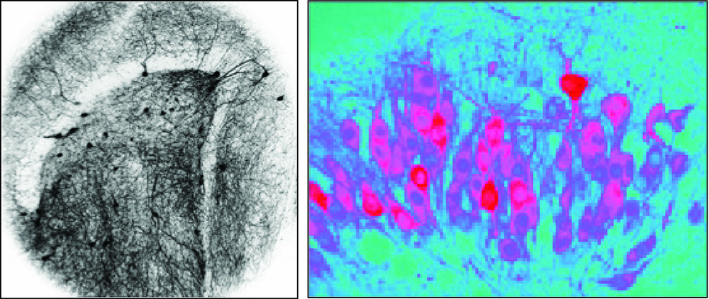
Left Image: Interneuronal network in the dentate gyrus captured by two-photon excitation fluorescence microscopy. Image Courtesy of Hajime Takano, Christopher Dengler, Ethan Goldberg, Douglas Coulter (Translational Epilepsy Research Program) Right Image: Hippocampal neurons with the genetically encoded calcium indicator captured by two-photon excitation fluorescence microscopy. Image Courtesy of Hajime Takano, Fu-Chun Hsu, Douglas Coulter (Translational Epilepsy Research Program)
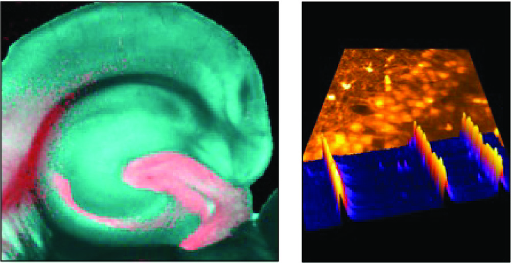
Left Image: Fluorescence micrograph of a hippocampal slice expressing red fluorescent protein in dentate gyrus granule cells. Image Courtesy of Hajime Takano and Douglas Coulter (Translational Epilepsy Research Program) Right Image: Fluorescence micrograph of CA3 hippocampal neurons stained with calcium indicator dye and color coded activity map of individual neurons captured by high speed confocal microscopy. Image Courtesy of Hajime Takano and Douglas Coulter (Translational Epilepsy Research Program)