HOW CAN WE HELP YOU? Call 1-800-TRY-CHOP
Precision Medicine Supports New Therapeutic Perspectives
Fine Control of Gene Therapy Products
Today’s gene therapies allow scientists to revise the script of a patient’s DNA to correct defective or deficient gene expression responsible for disease development. But once a therapy is delivered into a patient’s tissue, regulating levels of expression presents a challenge: Too much can have detrimental effects, while too little can fail to yield a result.
Children’s Hospital of Philadelphia researchers led by Beverly Davidson, PhD, director of the Raymond G. Perelman Center for Cellular and Molecular Therapeutics and chief scientific strategy officer at CHOP, engineered a delivery system — called the Xon system — to fine-tune levels of gene therapy expression, essentially a “dimmer switch” that advances precision medicine for common, rare, and complex conditions.
Together with collaborators from the Novartis Institutes for BioMedical Research (NIBR), Dr. Davidson and her team published the first research describing this innovation in Nature.
They showed that by using a small molecule drug in combination with gene therapy vectors, they could control dosing of protein expressed to achieve the maximum therapeutic benefit.
CHOP and NIBR are collaborating to develop next-generation small molecule splicing modulators and the Xon system to achieve fine-tuned gene regulation across multiple clinical applications.
Photomicrograph courtesy of Luis Tecedor
Computational Mining of Big Data
Combining computational mining of big data with experimental testing in the lab, researchers at Children’s Hospital of Philadelphia have identified RNA editing events that influence gene expression and, in turn, the phenotypic manifestation of that expression. In analyzing so-called A-to-I RNA editing, in which the adenosine of an RNA molecule is chemically modified into an inosine, the researchers describe how a single nucleotide change by RNA editing can have large downstream effects. The findings were published in Genome Biology.
Yi Xing, PhD, director of the Center for Computational and Genomic Medicine at CHOP and senior author of the study, worked with his team to study the functions of RNA editing through the lens of human genetic variation, or the differences that occur among people in approximately 1 in 1,000 DNA base pairs, affecting not only how genes are expressed but also how messenger RNAs are processed.
“What is so useful about this approach is that it is disease agnostic,” Dr. Xing said. “Future research can use this strategy to study specific diseases and look at the impact of RNA editing events from a disease perspective.”
Rebalancing Circuitries
A collaborative research team at Children’s Hospital of Philadelphia and the University of Pennsylvania found that a focal, molecular approach that targets circuit excitability may prove to be an effective intervention for treatment-resistant symptoms of mental illness.
Stewart Anderson, MD, associate chair of research, Department of Child and Adolescent Psychiatry and Behavioral Services, and associate director of the Lifespan Brain Institute at CHOP and the University of Pennsylvania, and Douglas Coulter, PhD, CHOP investigator and professor of Pediatrics at the Perelman School of Medicine at Penn, and colleagues published findings from their cross-disciplinary project in Biological Psychiatry.
The team identified circuits that normally underlie critical brain functions and compared them to instances of abnormal brain function in neurologic and psychiatric disease models to see if circuit disruptions could account for behavioral manifestations associated with the conditions. Then, they focally introduced “designer” receptors to correct the deficit.
The scientists identified hyperactive brain circuitry that affects social and spatial memory functions in a laboratory model of 22q11.2 deletion syndrome (22q11DS). They used focal gene therapy to correct the circuit hyperactivity and resolve the functional problem, thereby improving social and spatial memory. While translating this work from the lab to the clinic is a future goal, these findings are uniquely relevant to the 22q and You Center at CHOP for children who have hyperexcitability of the same hippocampal circuit.
Fascinating Insights into SARS-CoV-2
Children’s Hospital of Philadelphia researchers Paul Planet, MD, PhD, and Ahmed Moustafa, PhD, created a novel technology that traces the evolution of SARS CoV-2, called GNU-based Virus Identification (GNUVID). Drs. Planet and Moustafa gathered new findings that shed light on the importance of mask mandate policies and the effect of human immunity on viral diversity. With enhanced understanding of viral diversity, scientists are better positioned to detect concerning mutations and develop effective approaches to treatment and prevention.
GNUVID is an automatic tool that can take all currently available data and systematically name different viruses in the data bank, then sequence and deposit them in a database called GISAID that enables researchers to see where each virus comes from, where was it seen last, and, perhaps in the future, will allow them to learn which person it came from as the virus mutates. Noted by the scientists as the ultimate contact tracing, this tool can distinguish different viruses rapidly to see where they were transmitted from.
Dr. Moustafa, lead scientist in the Microbial Archive and Cryo-collection unit under the PennCHOP Microbiome Program, identified the dates in which mask mandates were put into place for 16 states in the United States to learn about the circulating diversity in those states over the period of the pandemic. Using GNUVID, Drs. Planet and Moustafa can categorize and follow new mutants as they arise, and then track those pathogens back to their closest relatives and to others circulating around the world and in the Philadelphia community.
Clearer Picture of Lymphatic System at Work
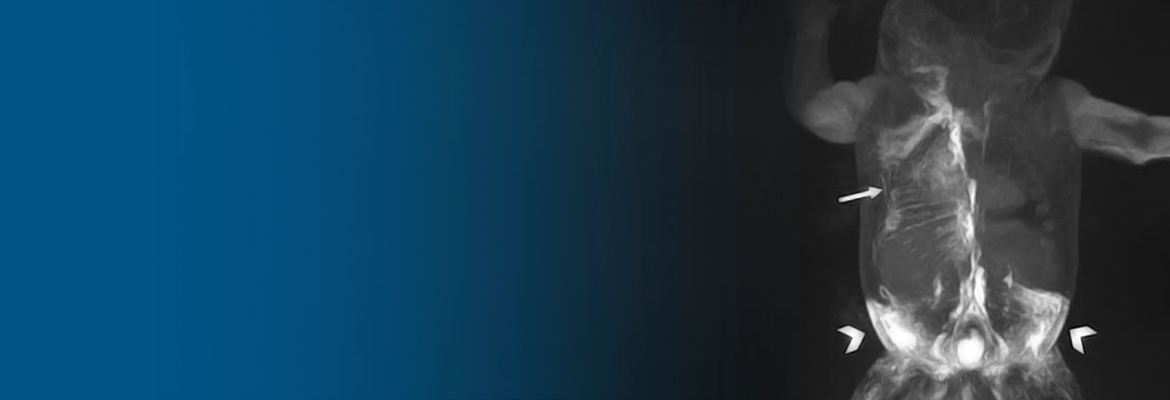
Novel imaging methods developed by Children’s Hospital of Philadelphia researchers help clinicians have a clearer picture of the lymphatic system at work — an innovation that has led to improved outcomes for infants with severe neonatal lymphatic disorders.
Researchers in the Jill and Mark Fishman Center for Lymphatic Disorders described the impact of these novel, minimally invasive imaging methods on two complex neonatal lymphatic disorders: neonatal chylothorax (NCTx) and central lymphatic flow disorder (CLFD) in a paper that appeared in the Journal of Perinatology. With high mortality and morbidity rates, detecting and diagnosing these disorders early is critical, in order to direct the best treatment strategies.
“NCTx and CLFD have been historically challenging to diagnose because there really wasn’t a way to image the system,” said Erin Pinto, MSN, RN, CCRN, first author of the paper and a nurse practitioner in the Center for Lymphatic Disorders.
Yoav Dori, MD, PhD, director of Pediatric Lymphatic Imaging and Interventions and Lymphatic Research, developed a method of accessing different compartments of the lymphatic system called dynamic contrast MR lymphangiography (DCMRL) that essentially “maps” the anatomy of a patient’s lymphatic system.
High-resolution 3D Structural Information for Proteins
Data generated using nuclear magnetic resonance (NMR) spectroscopy holds great promise, but researchers are often frustrated by how laborious it can be to perform the analysis before they can begin to derive meaningful results.
Nikolaos Sgourakis, PhD, lead principal investigator in the Mechanistic Molecular Immunology Lab at Children’s Hospital of Philadelphia, with colleagues from the University of California Santa Cruz and Universidade Federal do Rio de Janeiro, developed a new procedure for recording and analyzing NMR data, methyl assignments using satisfiability (MAUS), that effectively tackles these challenges. Now, solving a problem that would normally take weeks of NMR machine and expert time can be accomplished in just a few days.
MAUS allows biomedical researchers to visualize protein molecules with atomic-detail accuracy, opening new possibilities in drug development. With MAUS, researchers can apply the analysis method to larger, more complex proteins to understand their interactions with other molecules and induced structural changes — both aspects that are critical for targeting proteins often deemed undruggable because their predominant structures offer limited surfaces for interactions with antibodies and small molecules.
Their paper in Nature Communications demonstrates the effectiveness of MAUS on a range of important protein molecules, including a common form of the human leucocyte antigen, the therapeutic cytokine interleukin 2, and the Cas9 nuclease.


