HOW CAN WE HELP YOU? Call 1-800-TRY-CHOP

Scientists Make Big Strides in Research that Spans a Lifetime

The best CPR approaches for children are not necessarily identical to those for adults. This year, CHOP researchers published findings in the Journal of the American College of Cardiology that support the use of bystander CPR with rescue breathing in children experiencing cardiac arrest. The finding is a departure from the compression-only CPR recommended for adults by the American Heart Association (AHA) and the European Resuscitation Council (ERC).
While compression-only CPR has shown to be as effective as CPR with rescue breathing in adults, pediatric cardiac arrest often stems from breathing problems. Thus researchers at CHOP hypothesized the former approach might actually prove less effective in children.
“At the moment, most lay people are trained in compression-only CPR because that is the standard of care in adults,” said Maryam Y. Naim, MD, MSCE, a pediatric cardiac intensive care physician in the Division of Cardiac Critical Care Medicine and first author of the study. “However, children are not simply small adults, and our study shows there is a tremendous need for education in all communities about the benefits of CPR with rescue breathing in the pediatric population.”
Using data from the Cardiac Arrest Registry to Enhance Survival (CARES) database, Dr. Naim and her team analyzed more than 10,000 out-of-hospital cardiac arrests in patients between 0 and 18 years of age. They found that less than half of those who experienced pediatric cardiac arrest outside of the hospital received bystander CPR and of those who did, the majority received compression-only CPR. Compared to those who received compression-only CPR, children who received CPR with rescue breathing were nearly 1.5 times as likely to have better neurological outcomes. Further supporting the use of rescue breathing, the researchers found that infants who received compression-only CPR had the same outcomes as infants who received no CPR at all.
The results carry critical implications for bystander CPR education and training, including the need to emphasize and teach lay rescuers how to perform rescue breathing, according to Dr. Naim.

Critical period of bone growth
The discovery of dozens of new genetic markers associated with bone mineral accrual has set the stage for using genetic testing to identify the causes of eventual osteoporosis.
Publishing their findings in Genome Biology, the multidisciplinary team of genetics and bone biology experts at CHOP used data from the Bone Mineral Density in Childhood Study (BMDCS) to follow a group of children over several years.
“We wanted to do a GWAS study that measured bone mineral accrual at multiple time points to provide us with proper longitudinal data at ages when the skeleton is growing and developing,” said Struan F.A. Grant, PhD, director of the Center for Spatial and Functional Genomics and lead author of the study. “By doing a longitudinal study, we had much greater power in a relatively small cohort of patients.”
Accruing bone density in childhood is critical for achieving optimal bone mass as an adult. Until now, however, few studies have looked at genetic markers for bone health during this important period of growth. In the study, Dr. Grant and his team identified 40 distinct loci, or genetic markers, associated with bone accrual, several of which are associated with later fracture risk. The team also identified two novel, potentially causative effector genes, as well as multiple genetic pathways involved in bone accrual variation that help determine whether cells eventually become osteoblasts (bone cells) or adipocytes (fat cells).
According to Babette Zemel, PhD, the study’s first author and associate program director of CHOP’s Center for Human Phenomic Science, the findings demonstrate how such genetic markers manifest themselves earlier in life than previously thought. “In this case, our findings may help us better tailor lifestyle interventions, such as exercise and dietary changes, that will help patients later in life, and they may also lead to novel therapeutic interventions,” Dr. Zemel said.

Next Generation of Fetal Medicine Breakthroughs
A transformational gift of $25 million from Richard Wood Jr., the chairperson emeritus of Wawa, allows Children’s Hospital of Philadelphia scientists to continue advancing fetal medicine research and treatment for the next generation of tiny patients. In recognition of the donation, which was gifted in the spring of FY2020, CHOP re-named the Center for Fetal Diagnosis and Treatment (CFDT) the Richard D. Wood Center for Fetal Diagnosis and Treatment.
First opened in 1995, the internationally recognized CFDT is home to groundbreaking scientific breakthroughs, most notably the research, development, and accomplishment of the first successful fetal surgical repair of spina bifida. The Center’s director, N. Scott Adzick, MD, and its leaders are considered founders of modern fetal medicine for providing life-changing surgical options for unborn patients with spina bifida, congenital diaphragmatic hernia, Twin-Twin Transfusion Syndrome, lung lesions, sacrococcygeal teratoma, and lower urinary tract obstruction.
The new donation will allow Dr. Adzick and his team to expand their clinical, educational, and research efforts even further, including the construction of a new clinical space for the Garbose Family Special Delivery Unit, the creation of a birth defects biorepository, and the establishment of endowments for a Distinguished Chair in Pediatric Surgical Science as well as fellowships in Pediatric Surgical Science.
“It’s a great privilege and honor to name the hospital’s Center for Fetal Diagnosis and Treatment after Richard D. Wood Jr.,” said Dr. Adzick, who is also Surgeon-in-Chief of CHOP’s Department of Surgery. “On behalf of our entire team, I would like to express my gratitude to the Wood family on this historic gift, which will fuel a new era of breakthroughs in fetal medicine and surgery. Their generous philanthropic support will allow for a major expansion of infrastructure, patient services, research, and recruitment that will categorically be pivotal to our hospital and the patients and families we serve worldwide.”

Creating a world-class resourceREATING A WORLD-CLASS RESOURCE
A new data coordinating center (DCC) co-led by CHOP scientists seeks to improve the lives of individuals with Down syndrome by accelerating research and advancing medical care.
Supported by a five-year $19.5 million grant from the National Institutes of Health’s Investigation of Co-occurring conditions across the Lifespan to Understand Down syndrome (INCLUDE) project, our Center for Data Driven Discovery in Biomedicine (D3b) is developing the DCC in partnership with Sage Bionetworks and the Linda Crnic Institute for Down Syndrome at the University of Colorado Anschutz Medical Campus.
The project’s leaders, including our own Adam Resnick, PhD, envision the INCLUDE DCC as a world-class resource for data sharing, data access, and integrative analysis in Down syndrome. Three specialized cores — the Data Portal, Data Management, and Administration and Outreach — will work together to advance scientists’ understanding of Down syndrome, including the biology behind why individuals living with the condition have an increased risk for some medical conditions (such as hearing loss) but not others (such as heart disease).
“More and more, the scientific community is demonstrating the power of platforms to connect different communities with diverse areas of expertise and datasets to drive surprising discoveries and accelerated impact across a broad number of conditions in both children and adults,” said Dr. Resnick, who is D3b’s director. “The DCC Project will build on these efforts through the implementation of new technologies and platforms that will empower large-scale, diverse INCLUDE datasets on behalf of individuals with Down syndrome and other associated medical conditions and diseases.”

Culmination of TODAY2 Study
Treating type 2 diabetes in youth at an earlier point in time is critical, according to novel findings from CHOP and Penn researchers. In a landmark study published in the New England Journal of Medicine, the researchers reported that young people diagnosed with type 2 diabetes during childhood and adolescence have a high risk of developing serious complications by early adulthood. The complications, which can include high blood pressure, kidney disease, and others, accumulate rapidly: 60 percent of study participants developed at least one condition by early adulthood. Nearly a third of participants had two or more complications.
The Treatment Options for Type 2 Diabetes in Adolescents and Youth (TODAY) follow-up study (TODAY2) monitored 500 patients from the original TODAY study every year for signs of diabetes complications, from heart disease to kidney disease to diabetic foot complications, and more. When the participants enrolled, they ranged between the ages of 10 and 17, and had been diagnosed with type 2 diabetes for fewer than two years. The TODAY2 study found that 67 percent of participants developed high blood pressure, nearly 55 percent had kidney disease, and 51 percent had eye disease. Almost a third of patients showed signs of nerve disease.
“The results of this important study show that type 2 diabetes in children is a severe disease — more severe than in adults — and so we need to treat the disease aggressively at an earlier point on the timeline, ideally at the pre-diabetes stage,” said Lorraine Katz, MD, director of the Center for Human Phenomic Science at CHOP and PI of the study’s CHOP site. “We also must explore better therapeutic options for young people with type 2 diabetes to prevent the disease from worsening and leading to serious complications.”
Myelination's Key Roles in Life Stages
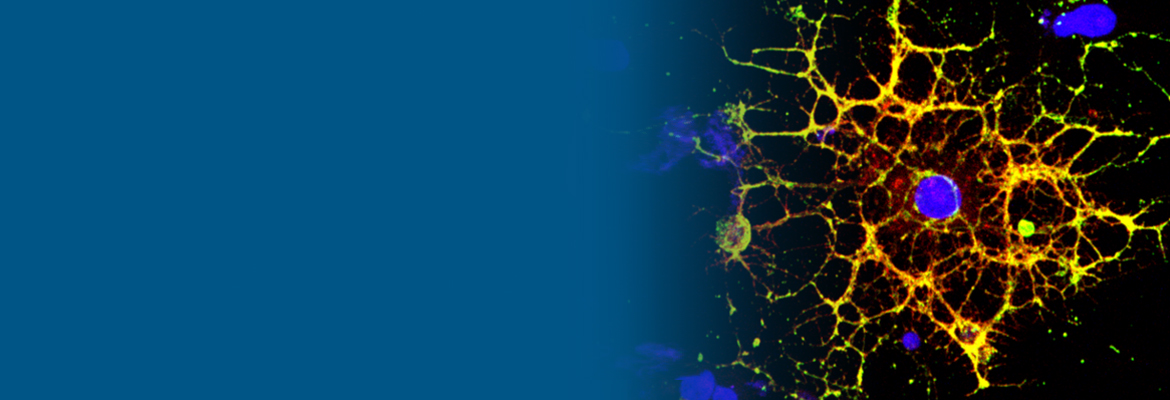
While it’s well known that individuals living with HIV experience a loss of white matter in the brain, not much is known about how exactly the virus contributes to such reduction. This year, researchers at CHOP and Penn described in Glia the mechanism by which HIV infection blocks the maturation process of the brain cells that produce myelin, the substance white matter is made of that provides neurons with a protective coat.
“When people think about the brain, they think of neurons, but they often don’t think about white matter, as important as it is,” said Judith B. Grinspan, PhD, a research scientist at CHOP. “But it’s clear that myelination is playing key roles in various stages of life: in infancy, in adolescence, and likely during learning in adulthood too. The more we find out about this biology, the more we can do to prevent white matter loss and the harms that can cause.”
By looking at both human and animal cells for the study, Dr. Grinspan and her team were able to detail how HIV hinders the maturation of oligodendrocytes, the brain cells that make myelin, and thus reduce the production of white matter. Testing the finding further, the team applied a compound that blocks this process and found that oligodendrocytes could once again mature. The findings are key for discerning how much of the white matter loss experienced by patients with HIV can be attributed to the virus itself, and how much can be attributed to antiretroviral therapy (ART), drugs used to treat HIV that have been shown by the team to also cause myelin reduction.
“When we put people on ART, especially kids or adolescents, it’s important to understand the implications of doing that,” said Kelly Jordan-Sciutto, PhD, Dr. Grinspan’s collaborator and a professor in Penn’s School of Dental Medicine, in Penn Today. “Antiretrovirals may prevent the establishment of a viral reservoir in the central nervous system, which would be wonderful, but we also know that the drugs can cause harm, particularly to white matter.
Image courtesy of Raj Putatunda.